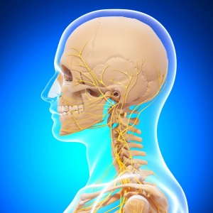
Over the last decade, research has demonstrated that spinal manipulation can change various aspects of nervous system function, including muscle reflexes, cognitive processing, reaction time, and the speed at which the brain processes information. One research group from New Zealand (Haavik et al) has hypothesized that the articular dysfunction part of the chiropractic clinical construct, the vertebral subluxation, results in altered afferent input to the central nervous system (CNS) that modifies the way in which the CNS processes and integrates all subsequent sensory input. This processing (i.e., sensorimotor integration) is a central nervous system (CNS) function that appears most vulnerable to altered inputs.
Investigators utilizing techniques such as transcranial magnetic stimulation and somatosensory evoked electroencephalographic (EEG) potentials have suggested that neuroplastic changes occur in the brain (e.g. primary sensory cortex, primary motor cortex, prefrontal cortex, basal ganglia, and cerebellum). Inducing and recording somatosensory evoked potentials (SEPs) is emerging in scientific literature relating to spinal manipulation (SM). There is evidence to support that SEPs are able to elucidate differences in cortical activity associated with SM. Studies with only a few recording EEG electrodes allow investigation of evoked potential amplitudes and latencies and have shown changes in the N30 somatosensory evoked potential (SEP) amplitudes following spinal manipulation. The N30 response from the frontal lobe peak reflects sensory integration.
With recent advances in the spatial resolution of EEG, it is becoming possible to better anatomically localize the signal. With this study, the authors aimed to utilize brain electrical source analysis to explore which brain sources are responsible for changes in N30 amplitude following a single session of spinal manipulation.
Nineteen young (average age 26 years) subclinical pain volunteers were included in the study. Subclinical pain (SCP) refers to recurrent spinal ache, pain, or stiffness for which the subject had not sought treatment. Subjects were excluded if they had: no evidence of spinal dysfunction, they were in current pain, they had sought previous treatment for their spinal issues, or they had contraindications to receiving spinal manipulation. The EEG signals were recorded with the Neuroscan System from 62 scalp electrodes using the extended 10-20 system montage. Supine subjects received electrical stimulations applied to the median nerve at the right wrist to evoke SEPs. Two trials of 1000 pulses were given in each session: one trial before treatment (control or chiropractic) and one trial after the treatment.
The entire spine and both sacroiliac joints were assessed for segmental dysfunction and adjusted where they were deemed necessary by an experienced chiropractor. Assessment for dysfunction included tenderness to palpation of the relevant joints, restricted intersegmental range of motion, asymmetric muscle tension, and any abnormal or blocked joint play and end-feel of the joints. The control (sham) involved one of the investigators (not a chiropractor) simulating a chiropractic treatment session. This included passive and active movements of the subject’s head, spine, and body, similar to what was done by the chiropractor who provided the actual chiropractic treatment.
Results:
- SEPs were successfully recorded in all subjects
- the majority of subjects were able to correctly guess which intervention group they were in (SM or sham)
- there was a significant post-intervention difference between the two groups – specifically the N30 amplitude was reduced in the spinal manipulation group following the treatment, while it remained stable in the control group
- source localization indicated that the prefrontal cortex tended to have the highest strength during the time interval between 20 and 60 ms
- source strength analysis revealed that chiropractic treatment reduced the strength of the prefrontal source, while all the other strengths remained stable
Key Points:
- Results from this study confirmed that spinal manipulation of dysfunctional spinal segments reduces the N30 SEP peak amplitude and demonstrated that this change is taking place in the prefrontal cortex
- This suggests that, at least in part, the mechanisms by which spinal manipulation improves performance are due to a change in function at the prefrontal cortex
- It is possible that the mechanisms behind pain relief following spinal manipulation in low level pain patients are due to improved sensorimotor integration and appropriate motor control, as this is the key function of the prefrontal cortex
Source: Lelic D, Niazi IK, Holt K, Jochumsen M, Dremstrup K, Yielder P, Murphy B, Drewes AM, Haavik H. Manipulation of Dysfunctional Spinal Joints Affects Sensorimotor Integration in the Prefrontal Cortex: A Brain Source Localization Study. Neural Plast. 2016;2016:3704964.
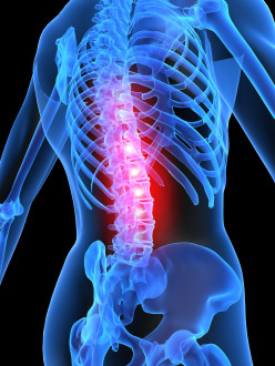
 Subacute and chronic patients with MRI confirmed symptomatic disc herniation treated with spinal manipulation were statistically (and clinically) significantly more likely to report improvement at 3 months compared with the nerve root injection. This prospective cohort study had 104 patients, 52 patients treated with cervical nerve root injection (CNRI) and 52 patients treated with spinal manipulation by a chiropractor. Baseline numerical rating scale (NRS) pain data were collected. Three months after treatment, numerical rating score pain levels were recorded and overall “improvement” was assessed using the Patient Global Impression of Change scale. Responses that were “much better” or “better” were considered to be “improved.” The proportion of patients “improved” was calculated for each treatment method and compared. The NRS and NRS change scores for the 2 groups were compared at baseline and 3 months. Results showed that there was no significant difference in outcomes between acute patients treated with cervical nerve root blocks and those treated with spinal manipulation at 3 months. However, when comparing the 3-month outcomes for the subacute/chronic patients, more than 78% of patients treated with SMT reported clinically relevant improvement compared with 37.5% of patients receiving a single CNRI. There were no adverse events for patients in either treatment group and the cost of treatment was similar for the 2 treatment procedures.
 Over the last decade, research has demonstrated that spinal manipulation can change various aspects of nervous system function, including muscle reflexes, cognitive processing, reaction time, and the speed at which the brain processes information. One research group from New Zealand (Haavik et al) has hypothesized that the articular dysfunction part of the chiropractic clinical construct, the vertebral subluxation, results in altered afferent input to the central nervous system (CNS) that modifies the way in which the CNS processes and integrates all subsequent sensory input. This processing (i.e., sensorimotor integration) is a central nervous system (CNS) function that appears most vulnerable to altered inputs.
 Research on chiropractic spinal manipulation (CSM) has been conducted extensively worldwide, and its efficacy on musculoskeletal symptoms has been well documented. Previous studies have documented potential relationships between spinal dysfunction and the autonomic nervous system and that chiropractic treatment affects the autonomic nervous system.