shoulder pain manual therapy
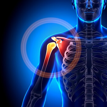
Shoulder pain is one of the most common musculoskeletal disorders. The lifetime prevalence is estimated to be in the range of 6.7–66.7%. Shoulder pain and stiffness may reduce family life or social life functions as well as reduce productive activities. It also has a strong statistical correlation with somatizing tendency and poor mental health. There are many cases of shoulder pain that have not improved over time, remain persistent, or occur repeatedly. The prognosis becomes poorer the longer the illness is present.  A review of the effectiveness of conservative nondrug, nonsurgical interventions, either alone or in combination, for conditions of the shoulder was published in the Journal of Manipulative and Physiological Therapeutics in June, 2017. Shoulder conditions addressed in the article were shoulder impingement syndrome (SIS), rotator cuff-associated disorders (RCs), adhesive capsulitis (AC), and nonspecific shoulder pain. Eligibility criteria for the scientific studies included randomized controlled trials (RCTs), systematic reviews, or meta-analyses. Treatments included nondrug, nonsurgical procedures. Results indicated low- to moderate-quality evidence supporting the use of manual therapies for all 4 shoulder conditions. Exercise, particularly combined with physical therapy protocols, was beneficial for SIS and AC. For SIS, moderate evidence supported several passive modalities. For RC, physical therapy protocols were found beneficial but not superior to surgery in the long term. Moderate evidence supported extracorporeal shockwave therapy for calcific tendinitis RC. Low-level laser was the only modality for which there was moderate evidence supporting its use for all 4 conditions.

Bottom line:

  • Manual therapy is beneficial for common shoulder conditions.
  • Low-level laser therapy is beneficial for common shoulder conditions.
  • Exercise protocols are beneficial for SIS and AC.


chiropractic neck pain whiplash
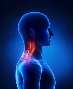
A clinical practice guideline on the management of neck pain–associated disorders (NADs) and whiplash-associated disorders (WADs) was recently developed and replaces existing chiropractic guidelines on these topics (Bussières, Stewart et al, 2016). The Guideline Development Group of the Canadian Chiropractic Guideline Initiative (CCGI) conducted the updated guidelines. They considered recently published systematic reviews on NAD and WAD from the Ontario Protocol for Traffic Injury Management (OPTIMa) Collaboration. Below is a brief summary of the guidelines.  Please refer to the numerous links in this blog post to gain access to the original paper and the full guidelines which are freely available.

Neck pain and its associated disorders (NAD), including headache and radiating pain into the arm and upper back, are common. These disorders often result in significant social, psychological, and economic burden.  Neck pain is a common reason for people to seek chiropractic care.

Motor vehicle collisions most commonly are associated with neck pain related to whiplash-associated disorders (WADs). Whiplash-associated disorders also affect the daily functioning of our patients in terms of considerable pain, suffering, disability, and costs.  Whiplash-associated disorders are defined as an injury to the neck that occurs with sudden acceleration or deceleration of the head and neck relative to other body parts.  Symptoms of WADs commonly include headache, stiffness, shoulder and back pain, numbness, dizziness, sleeping difficulties, fatigue, and cognitive deficits.

The 2000-2010 Bone and Joint Decade Task Force on Neck Pain and its Associated Disorders recommended that all types of neck pain, including WADs, be included under the classification of NAD.  The 4 grades of NAD are:

  • I – No signs or symptoms suggestive of major structural pathology and no or minor interference with activities of daily living
  • II – No signs or symptoms of major structural pathology, but major interference with activities of daily living
  • III – No signs or symptoms of major structural pathology, but presence of neurologic signs such as decreased deep tendon reflexes, weakness or sensory deficits
  • IV – Signs or symptoms of major structural pathology (e.g., fracture, tumor, infection)

After searching and synthesizing the latest scientific literature on these topics, the guideline committee provided their recommendations.  Below is the summary of the recommendations.  The full guideline and accompanying documents are available from the CCGI website at www.chiroguidelines.org. There are excellent resources for practitioners and patients available from this website including exercise videos and forms.

Global summary of recommendations: A multimodal approach including manual therapy, self-management advice and exercise is an effective treatment strategy for both recent onset and persistent neck pain and whiplash associated disorders.

A) Summary of Recommendations for Grades I-III Neck Pain and Associated Disorders (NAD)

  • For recent-onset (0-3 months) neck pain grades I-II, based on patient preference and practitioner experience we suggest offering advice with:
    • multimodal care;
    • manipulation or mobilization;
    • Range of motion home exercises or multimodal manual therapy.
  • For recent-onset (0-3 months) neck pain grade III, based on patient preference and practitioner experience we suggest offering advice with:
    • supervised graded strengthening exercises.
  • For persistent (>3 months) neck pain grades I-II, based on patient preference and practitioner experience we suggest offering advice with:
    • multimodal care or stress self-management;
    • multimodal care or advice alone;
    • manipulation in conjunction with soft tissue therapy;
    • supervised yoga; supervised group exercise; supervised strengthening exercises or home exercises;
    • mixed supervised and unsupervised high-intensity strength training or advice alone for workers with persistent neck and shoulder pain;
    • high dose massage.
  • For persistent (>3 months) neck pain grade III, based on patient preference and practitioner experience we suggest offering advice with:
    • multimodal care or advice alone;
    • mixed supervised and unsupervised high-intensity strength training or advice alone for workers with persistent neck and shoulder pain.

B) Summary of Recommendations for Grade I-III Whiplash and Associated Disorders (WAD)

  • For recent onset (0-3 months) whiplash grades I-III, based on patient preference and practitioner experience we suggest offering advice with:
    • multimodal care.
  • For persistent (>3 months) whiplash grades I-II, based on patient preference and practitioner experience we suggest offering advice with:
    • supervised exercise or advice alone.

Source: Bussières AE, Stewart G, Al-Zoubi F et al. The Treatment of Neck Pain-Associated Disorders and Whiplash-Associated Disorders: A Clinical Practice Guideline. J Manipulative Physiol Ther. 2016 Oct;39(8):523-564.

In addition to the profession’s largest presentation of research abstracts, DC2017 has added a Researcher’s Forum for Saturday, March 18 that will focus on funding opportunities from around the globe followed by a general discussion of all things research. See you there! https://www.acatoday.org/DC2017

1400 Introduction – WFC Research Council Chair and Vice-Chair
1410 Funding opportunities around the globe
NCCIH, USA – Wendy Weber or delegate (10 min total)
ECCRE, Europe – Henrik Wulff Christensen or delegate (10 min total)
CCRF, Canada – Alison Dantas / Ronda Parks or delegate (10 min total)
CAA, Australia – Scott Charlton or delegate (10 min total)
COCA, Australia – Simon French or delegate (10 min total)
CRC, England – Elisabeth Angier or delegate – (10 min total)
Crowd-Sourced Funding – Greg Kawchuk – (10 min total)
Questions – (10 min)

Break 15:30 – 16:00

16:00 Open Research Forum (all) – WFC Research Council Chair and Vice-Chair
An open forum for all attendees to discuss research topics related to the profession
1730 – End


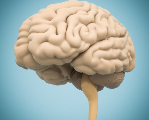
Chiropractic care is commonly thought to have a beneficial effect on the functioning of the human body by affecting the nervous system. Evidence indicates that chiropractic adjustments result in plastic changes in sensorimotor integration within the central nervous system in human participants, particularly within the prefrontal cortex. Adjustments appear to alter the net excitability of the low-threshold motor units, increase cortical drive, and prevent fatigue (see this blog).  This same group of researchers have more recently found an increase cortical drive to upper and lower extremity muscles following manipulation as measured by motor evoked potential. The researchers suggested the effects were due to descending cortical drive and could not be explained by changes at the level of the spinal cord (although they can’t rule that out completely).  Two experiments were conducted.  In experiment one, transcranial magnetic stimulation input–output (TMS I/O) curves for an upper limb muscle (abductor pollicus brevis; APB) were recorded, along with F waves prior to and after either spinal manipulation or a control intervention for the same subjects on two different days. During these two separate days, lower limb TMS I/O curves and movement related cortical potentials (MRCPs) were recorded from tibialis anterior muscle (TA) before and after spinal manipulation. Spinal manipulation resulted in a 54.5% ± 93.1% increase in maximum motor evoked potential (MEPmax) for APB and a 44.6% ± 69.6% increase in MEPmax for TA. 
They conclude that “Spinal manipulation may therefore be indicated for the patients who have lost tonus of their muscle and or are recovering from muscle degrading dysfunctions such as stroke or orthopaedic operations. These results may also be of interest to sports performers. We suggest these findings should be followed up in the relevant populations.”

Reference: Haavik H, Niazi IK, Jochumsen M, Sherwin D, Flavel S, Türker KS. Impact of Spinal Manipulation on Cortical Drive to Upper and Lower Limb Muscles. Brain Sci. 2016 Dec 23;7(1).


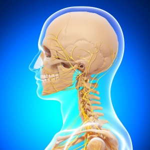
Subacute and chronic patients with MRI confirmed symptomatic disc herniation treated with spinal manipulation were statistically (and clinically) significantly more likely to report improvement at 3 months compared with the nerve root injection. This prospective cohort study had 104 patients, 52 patients treated with cervical nerve root injection (CNRI) and 52 patients treated with spinal manipulation by a chiropractor. Baseline numerical rating scale (NRS) pain data were collected. Three months after treatment, numerical rating score pain levels were recorded and overall “improvement” was assessed using the Patient Global Impression of Change scale. Responses that were “much better” or “better” were considered to be “improved.” The proportion of patients “improved” was calculated for each treatment method and compared. The NRS and NRS change scores for the 2 groups were compared at baseline and 3 months.  Results showed that there was no significant difference in outcomes between acute patients treated with cervical nerve root blocks and those treated with spinal manipulation at 3 months. However, when comparing the 3-month outcomes for the subacute/chronic patients, more than 78% of patients treated with SMT reported clinically relevant improvement compared with 37.5% of patients receiving a single CNRI. There were no adverse events for patients in either treatment group and the cost of treatment was similar for the 2 treatment procedures.

Reference: Peterson CK, Pfirrmann CW, Hodler J, Leemann S, Schmid C, Anklin B, Humphreys
BK. Symptomatic, Magnetic Resonance Imaging-Confirmed Cervical Disk Herniation Patients: A Comparative-Effectiveness Prospective Observational Study of 2 Age- and Sex-Matched Cohorts Treated With Either Imaging-Guided Indirect Cervical Nerve Root Injections or Spinal Manipulative Therapy. J Manipulative Physiol Ther. 2016 Mar-Apr;39(3):210-7.

Thanks for listening to the chiropractic science podcast this year.  Here are the top 5 downloads for 2016.  Happy holidays! Look for more great speakers in 2017!

Dr. Scott Haldeman - Pioneering Chiropractic Research, Chiropractic Science

1. Dr. Scott Haldeman

DrJanHartvigsenthumbnail

2. Dr. Jan Hartvigsen

Heidi_Haavik_web

3. Dr. Heidi Haavik

gregthumbnail

4. Dr. Greg Kawchuk

Dr. Lise Hestbaek

5. Dr. Lise Hestbæk


adobestock_49611047
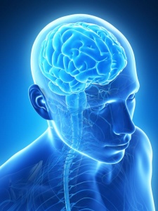
Over the last decade, research has demonstrated that spinal manipulation can change various aspects of nervous system function, including muscle reflexes, cognitive processing, reaction time, and the speed at which the brain processes information. One research group from New Zealand (Haavik et al) has hypothesized that the articular dysfunction part of the chiropractic clinical construct, the vertebral subluxation, results in altered afferent input to the central nervous system (CNS) that modifies the way in which the CNS processes and integrates all subsequent sensory input. This processing (i.e., sensorimotor integration) is a central nervous system (CNS) function that appears most vulnerable to altered inputs.

Investigators utilizing techniques such as transcranial magnetic stimulation and somatosensory evoked electroencephalographic (EEG) potentials have suggested that neuroplastic changes occur in the brain (e.g. primary sensory cortex, primary motor cortex, prefrontal cortex, basal ganglia, and cerebellum).  Inducing and recording somatosensory evoked potentials (SEPs) is emerging in scientific literature relating to spinal manipulation (SM). There is evidence to support that SEPs are able to elucidate differences in cortical activity associated with SM. Studies with only a few recording EEG electrodes allow investigation of evoked potential amplitudes and latencies and have shown changes in the N30 somatosensory evoked potential (SEP) amplitudes following spinal manipulation.  The N30 response from the frontal lobe peak reflects sensory integration.

With recent advances in the spatial resolution of EEG, it is becoming possible to better anatomically localize the signal.  With this study, the authors aimed to utilize brain electrical source analysis to explore which brain sources are responsible for changes in N30 amplitude following a single session of spinal manipulation.

Nineteen young (average age 26 years) subclinical pain volunteers were included in the study. Subclinical pain (SCP) refers to recurrent spinal ache, pain, or stiffness for which the subject had not sought treatment. Subjects were excluded if they had: no evidence of spinal dysfunction, they were in current pain, they had sought previous treatment for their spinal issues, or they had contraindications to receiving spinal manipulation. The EEG signals were recorded with the Neuroscan System from 62 scalp electrodes using the extended 10-20 system montage. Supine subjects received electrical stimulations applied to the median nerve at the right wrist to evoke SEPs. Two trials of 1000 pulses were given in each session: one trial before treatment (control or chiropractic) and one trial after the treatment.

The entire spine and both sacroiliac joints were assessed for segmental dysfunction and adjusted where they were deemed necessary by an experienced chiropractor. Assessment for dysfunction included tenderness to palpation of the relevant joints, restricted intersegmental range of motion, asymmetric muscle tension, and any abnormal or blocked joint play and end-feel of the joints. The control (sham) involved one of the investigators (not a chiropractor) simulating a chiropractic treatment session. This included passive and active movements of the subject’s head, spine, and body, similar to what was done by the chiropractor who provided the actual chiropractic treatment.

Results:

  • SEPs were successfully recorded in all subjects
  • the majority of subjects were able to correctly guess which intervention group they were in (SM or sham)
  • there was a significant post-intervention difference between the two groups – specifically the N30 amplitude was reduced in the spinal manipulation group following the treatment, while it remained stable in the control group
  • source localization indicated that the prefrontal cortex tended to have the highest strength during the time interval between 20 and 60 ms
  • source strength analysis revealed that chiropractic treatment reduced the strength of the prefrontal source, while all the other strengths remained stable

Key Points:

  • Results from this study confirmed that spinal manipulation of dysfunctional spinal segments reduces the N30 SEP peak amplitude and demonstrated that this change is taking place in the prefrontal cortex
  • This suggests that, at least in part, the mechanisms by which spinal manipulation improves performance are due to a change in function at the prefrontal cortex
  • It is possible that the mechanisms behind pain relief following spinal manipulation in low level pain patients are due to improved sensorimotor integration and appropriate motor control, as this is the key function of the prefrontal cortex

Source: Lelic D, Niazi IK, Holt K, Jochumsen M, Dremstrup K, Yielder P, Murphy B, Drewes AM, Haavik H. Manipulation of Dysfunctional Spinal Joints Affects Sensorimotor Integration in the Prefrontal Cortex: A Brain Source Localization Study. Neural Plast. 2016;2016:3704964.


adobestock_50009138

The high prevalence of low-back pain (LBP) has been highlighted for many years, but until recently, awareness of its influence on the population was inadequate. The results of the Global Burden of Disease (GBD) Projects 2010 have informed us that the leading cause of disability (as measured by years lived with disability) worldwide is low back pain.  Additionally, musculoskeletal conditions as a whole are the second greatest cause of disability globally according to a report by international experts, published in The Lancet on December 15th, 2012.  In the first comprehensive study of the worldwide impact of all diseases and risk factors, musculoskeletal (MSK) conditions such as arthritis and back pain affect over 1.7 billion people worldwide, and have the fourth greatest impact on the overall health of the world population, considering both death and disability.  This burden has increased by 45% over the last 20 years and will continue to do so unless action is taken.  This landmark study of the global burden of all diseases provides indisputable evidence that musculoskeletal conditions are an enormous and emerging problem in all parts of the world and need to be given the same priority for policy and resources as other major conditions like cancer, mental health and cardiovascular disease.

With the knowledge that LBP is the number one cause of disability in the world, it is unfortunate that little is known about the detailed course, and trajectory, of LBP.  Until recently LBP was believed to be a self-limiting condition, similar to the common cold. However, research in the past two decades has shown that LBP is actually a recurrent condition that could be likened to a more chronic condition such as asthma.  In this regard, we are starting to look at LBP as not seen as a single entity, but rather to the LBP condition which can be regarded as a chain of LBP episodes.  So, we need to view LBP (and maybe all types of spine pain) as having a lifelong course – perhaps with different etiology and modifying factors as life progresses, but always existing as an underlying ‘trait’.

When researchers have looked at the non-benign, and non self-limiting nature of the condition, three large groups of LBP patients emerge: 1): those without LBP; 2) those who experience it on and off and; 3) those who have it most of the time. It is pretty clear that definite recovery with no recurrences does not appear to be common, although to date, we do not know how these patterns develop over the course of a lifetime. People with LBP will not necessarily seek care, but a person who consults a chiropractor for an episode of LBP is likely to feel better fairly quickly. In light of these findings, clinicians should observe and convey information about episodes within the context of a longer-term pain trajectory, to provide patients with a realistic view of the problem. The authors of the recent trajectories of low back pain article referenced herein suggest that effective short-term treatment strategies, pain management and activity maintenance as well as secondary and tertiary prevention should be high on the clinical agenda. ‘Management rather than cure’ might be a helpful catch phrase, similar to the well-known recommendation of ‘don’t worry – keep active’ (Axén and Leboeuf-Yde, 2013).

Given the shift in attention of LBP to view it as a chronic condition, researchers and clinicians are putting more emphasis on investigating LBP throughout the life course.  What is emerging from this life course investigation is that similar factors (e.g., genetics, parental factors, psychological factors, injury, physical activity, comorbidity) are associated with the pain at different times. There appears to be strong evidence for the links between back pain, pain at other locations (e.g., shoulder) and other health problems. This evidence leads to the potential conclusion that vulnerability for long-term back pain develops at an early age, likely in childhood, and influences the occurrence of, and recovery from, episodes of back pain (Dunn et al, 2013).

Furthermore, results of a recent meta-analysis of LBP in children and adolescents indicates higher prevalence rates of LBP in the most recent studies suggesting that this a problem that is increasing in this young population (Calvo-Muñoz et al, 2013). As a consequence, more attention should be devoted to develop and apply prevention programs and early detection programs for young children in order to reverse this tendency.

Key Points

  • The leading cause of disability worldwide is low back pain
  • Evidence is mounting that classifying low back pain as acute, subacute and chronic is no longer helpful
  • Many individuals experience multiple episodes of back pain with the first episode occurring early in life
  • LBP is now being thought of as a potentially chronic health condition in its own right

References:

1. Vos T et al.Years lived with disability (YLDs) for 1160 sequelae of 289 diseases and injuries 1990-2010: a systematic analysis for the Global Burden of Disease Study 2010. Lancet. 2012 Dec 15;380(9859):2163-96.

2.Axén I, Leboeuf-Yde C. Trajectories of low back pain. Best Pract Res Clin Rheumatol. 2013 Oct;27(5):601-12.

3. Dunn KM, Hestbaek L, Cassidy JD. Low back pain across the life course. Best Pract Res Clin Rheumatol. 2013 Oct;27(5):591-600.

4. Calvo-Muñoz I, Gómez-Conesa A, Sánchez-Meca J. Prevalence of low back pain in children and adolescents: a meta-analysis. BMC Pediatr. 2013 Jan 26;13:14.

adobestock_69723890
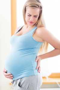
Low back pain is one of the most common and often disabling problems in pregnancy. The prevalence of pregnancy related low back pain (PLBP) or pelvic girdle pain (PGP) is 20% to 90% with most studies reporting more than 50% prevalence. —PGP is almost 2x more common than lumbar pain. —25% of all postpartum women suffer from PGP and/or PLBP.

A 2014 prospective, cohort, outcomes study involving 115 pregnant women with low back or pelvic pain participated in the study.  Baseline numerical rating scale (NRS) of pain intensity and Oswestry Low Back Pain Disability Index questionnaire data were collected.  In addition, The patient’s global impression of change (PGIC) (primary outcome), NRS, and Oswestry data (secondary outcomes) were collected at 1 week, 1 and 3 months after the first treatment.  Then, at 6 months and 1 year the PGIC and NRS scores were collected again. PGIC responses of ‘better’ or ‘much better’ were categorized as ‘improved’. Chiropractic treatment was pragmatic and left to the discretion of the treating clinician.

Results:

  • 52% of 115 recruited patients ‘improved’ at 1 week, 70% at 1 month, 85% at 3 months, 90% at 6 months and 88% at 1 year.
  • There were significant reductions in NRS and Oswestry scores
  • Patients with more prior LBP episodes had higher 1 year NRS scores

Most pregnant patients with low back or pelvic pain undergoing chiropractic treatment reported clinically relevant improvement at all time points.

Reference: Peterson CK, Mühlemann D, Humphreys BK. Outcomes of pregnant patients with low back pain undergoing chiropractic treatment: a prospective cohort study with short term, medium term and 1 year follow up. Chiropr Man Therap. 2014 Apr 1;22(1):15.


adobestock_42898239Infantile colic is one of the significant challenges of parenthood.  It is one of the common reasons for pediatrician visits during the child’s first 3 months of life. Infantile colic is a prevalent and distressing condition for which there is no proven standard therapy, which motivates parents to seek alternatives.  It is defined as paroxysms of crying lasting more than 3 hours a day, occurring more than 3 days in any week for 3 weeks (aka rule of 3) in a healthy baby aged 2 weeks to 4 months. Colic remains a poorly understood phenomenon affecting up to 30% of babies, with underlying organic causes of excessive crying accounting for less than 5% of cases.  Laboratory tests and radiological examinations are unnecessary if the infant is gaining weight normally and has a normal physical examination.

To date, several randomized trials examining chiropractic care for children with colic have been reported, and although these trials demonstrate some reduction in crying, weaknesses in study methodologies have limited the evidence they provide.  Based on these previous studies, there is some but not definitive evidence to make a recommendation of manual therapy for the excessively crying baby.

The purpose of this study was to try to address methodological weaknesses in the scientific literature by conducting a single-blind, randomized controlled trial comparing chiropractic manual therapy with no treatment and to determine whether parents’ knowledge of treatment biases their report of change in infant crying.

Infants with unexplained persistent crying (colic) verified by a baseline crying diary of 3 days or more and presenting to the Anglo-European College of Chiropractic were included in the study. Other inclusion criteria included: patients had to be younger than 8 weeks, born at a gestational age of 37 weeks or later, and had a birth weight of 2500 grams or more and show no signs of other conditions or illness.  One hundred and four infants participated.

Parents completed a questionnaire (baseline) and their child was then randomized to 1 of 3 groups.  In 2 of the 3 groups, infants received treatment, and in the third, no treatment was administered.  For one of the treatment groups, the parent was able to observe the treatment and knew that the infant was being treated.  Parents in the other two groups were seated behind a screen and could not observe their child. Therefore, parents in these two groups were ‘blind’ as to whether their infant received treatment or not.  To be clear, the 3 groups were: (i) infant treated/parent aware, (ii) infant treated/parent unaware (blinded), and (iii) infant not treated/parent unaware (blinded).

Chiropractic care was delivered by a chiropractic intern and involved low force tactile pressure to spinal joints and paraspinal muscles where dysfunction was noted on palpation. The manual therapy, estimated at 2 N of force, was given at the area of involvement without rotation of the spine. Treatment duration lasted up to 10 days, and the number of treatments during this period were influenced by examination findings and parent reports. Treatment was stopped if parents reported their infant was symptom-free. Infants in the blinded groups were placed by the parent on the examination table and then parents sat behind a screen that blinded observation. Patients in the no-treatment group were not touched by the intern and/or clinician.

Outcome measures included crying time as assessed by a 24 hour crying diary ending either 10 days after baseline or at discharge – whichever was sooner.  Crying time was extracted from the diaries.  A global improvement scale (GIS) was completed at either 10 days or discharge by parents and assessed their ratings of change since baseline (e.g., worse to much improvement).

Key findings of this study were:

  • Compared with baseline, by day 10, there was a significant decrease in crying time -44.4%,  51.2%, and 18.6% in the treatment groups ([Blinded] and [Not Blinded]) and the no-treatment group, respectively
  • In parents blinded to treatment allocation, using 2 or less hours of crying per day to determine a clinically significant improvement in crying time, the increased odds of improvement in treated infants compared with those not receiving treatment were statistically significant at day 8 (adjusted odds ratio [OR], 8.1) and at day 10 (adjusted OR, 11.8)
  • There was a similar greater odds of improvement with treatment compared with no treatment using the global improvement scale
  • The number needed to treat was 3 (indicating that 3 infants need to be treated to gain one additional improvement in crying time over no treatment)

In summary, this study found that excessively crying infants were at least 5 times less likely to cry if they were treated with chiropractic manual therapy than if they were not treated.  Infants who were treated were equally likely to improve, whether the parents were blinded to treatment or not.

Reference:  Miller JE, Newell D, Bolton JE. Efficacy of chiropractic manual therapy on infant colic: a pragmatic single-blind, randomized controlled trial. J Manipulative Physiol Ther. 2012 Oct;35(8):600-7.