ChiropracticScienceLogo1American chiropractors are very similar to chiropractors in other countries, as well as other health professionals in terms of their favorable attitudes towards evidence based practice (EBP) (1-2). The purpose of EBP is to promote effective chiropractic practice and enhance public health. Evidence based practice incorporates the best research evidence with individual clinical expertise and patient choice/values (3). There are however several limitations and barriers to implementing EBP that chiropractors express such as lack of research relevance, lack of time and insufficient skills for locating and appraising research (1). It is important to note that these same obstacles are also encountered by other professions including medicine and nursing.

In a recent survey, very few DCs indicated that computer, internet, or database access were barriers to their uptake of EBP (1). Since chiropractors are interested in implementing EBP, these findings underscore the importance of providing clinicians with training in EBP skills, particularly through online resources. It is likely that comprehensive and multi-faceted approaches that take into account all the relevant levels affecting EBP will likely be needed to integrate research into practice (1).

As a practitioner as well as a researcher, I realize the importance and barriers of translating research into practice. I have also begun to realize how I can give back to the profession. Some of you may recognize my On Target research summaries.  Others may be familiar with my chiropractic science podcast.  While there may be many ways to tackle the evidence-practice gap, one convenient educational method is a podcast. This is one of the ways that I have been utilizing to help get chiropractic research out to the masses. For those not familiar, a podcast describes audio and/or video files that can be downloaded and played on a personal computer or mobile device.

Podcast is the term commonly used to refer to a series of digital media files that are released periodically and can be subscribed to using an RSS (Really Simple Syndication) feed. This means of digital delivery is what makes podcasting different from other means of accessing media files over the internet.  Of course one can still download the audio and/or video files that makeup a podcast.  The automatically downloaded files can be stored locally on a computer or other device (such as your mobile phone) ready for offline use, providing simple and convenient access to regular releases.

Podcasting has been used successfully in teaching and learning in many different student groups (4-5). In theory podcasts provide educational content in a format that is convenient and available 24 hours a day and can be accessed whenever and wherever the learner chooses (4-5). Learners have the ability to replay a podcast as many times as they wish in order to improve understanding. In addition, podcasting offers the ability to embed additional content from researchers or clinicians to help make links between theoretical concepts and practice.

While many chiropractic podcasts exist, chiropractic science (CS) is the only podcast that exclusively interviews chiropractic researchers about the science of chiropractic. CS publicizes and disseminates chiropractic research. Hear about chiropractic research from the authors in plain English, not through the media, nor a middleman. Think about recent media coverage concerning the evidence of chiropractic in magazines or online.  While reports of chiropractic studies in the media may be very good (or very poor), some things get lost in translation.  By interviewing the chiropractic experts that are actually doing the research, you’ll get the information direct – the way the author intended it.

Making scientific findings available to the public is an important part of the research process.  Publicizing these interviews passes on the benefits of chiropractic research to other researchers, chiropractors in practice as well as practitioners from other disciplines and the wider community. In addition to publicizing the interviews, other goals of the podcast are to encourage collaboration of researchers to promote future high quality chiropractic research as well as to motivate and assist practitioners and students alike to pursue research careers in chiropractic science!

Why Chiropractic Science?

  • There is a lot of excellent chiropractic research published by top scientists in the field that deserve our attention
  • Incorporating the latest evidence into your practice of chiropractic is good for everyone
  • Reduce the evidence – practice gap and promote knowledge translation from the chiropractic research laboratory to the clinic
  • Engage clinicians in evidence based practice in a fun, easy to access manner
  • Learn from the experts and gain confidence in your knowledge and communications with others about chiropractic
  • A chiropractic resource for students, doctors and patients

Feedback about Chiropractic Science

  • “It has been such an inspiration to listen to all the greats in the world of chiropractic research”
  • “It has certainly made an impact on my practice and gives me a tremendous amount of certainty in what we do as chiropractors”
  • “I find the information profoundly motivating”
  • “It is difficult to read and interpret journal articles for a seasoned chiropractor. This saves time and hearing it explained from your colleagues themselves in language that I can communicate with my patients is invaluable”
  • “I am humbled by the sacrifice, hard work and accomplishments of all the researchers you have interviewed”
  • “Being in practice for many years, I am weary of ideologically biased forums when 21st Century chiropractic can have and bring it all; evidence and practice based science WITH a strong philosophical foundation. Highest accolades to Dr. Dean Smith for his untiring success at bringing the bar, raising colleagues and conversations together to articulate just how pertinent chiropractic is in contemporary health care”

In short, CS is starting to achieve what it set out to do, but this just the beginning. I am humbled by the feedback I have received from chiropractors about the podcast and am looking forward to interviewing as many chiropractic researchers as possible.  If you haven’t listened to the podcast, check them out.  If you like what you hear, please leave a great review on iTunes. This will help position CS as a leading health podcast – enlightening the masses about the evidence of chiropractic.  Listen to the podcast on iTunes or download the files directly to your device from chiropracticscience.com.  Enjoy and share with everyone.

References:

  1. Schneider MJ, Evans R, Haas M, Leach M, Hawk C, Long C, Cramer GD, Walters O, Vihstadt C, Terhorst L. US chiropractors’ attitudes, skills and use of evidence-based practice: A cross-sectional national survey. Chiropr Man Therap. 2015 May 4;23:16.
  2. Alcantara J, Leach MJ. Chiropractic Attitudes and Utilization of Evidence-Based Practice: The Use of the EBASE Questionnaire. Explore (NY). 2015 Sep-Oct;11(5):367-76.
  3. Lefebvre R, Peterson D, Haas M. Evidence-Based Practice and Chiropractic Care. J Evid Based Complementary Altern Med. 2012 Dec 28;18(1):75-79.
  4. Burke S, Cody W. Podcasting in undergraduate nursing programs. Nurse Educ. 2014 Sep-Oct;39(5):256-9.
  5. Strickland K, Gray C, Hill G. The use of podcasts to enhance research-teaching linkages in undergraduate nursing students. Nurse Educ Pract. 2012 Jul;12(4):210-4.


Dr. Dean Smith is founder of chiropractic science – dedicated to publicizing and disseminating chiropractic research through podcast interviews with the experts that are doing the research.  Listen to free chiropracticscience.com interviews on iTunes.  Dr. Smith is also a clinical professor at Miami University in Oxford, Ohio.  He has a practice of chiropractic in Eaton, Ohio.

Spinal manipulation biochemical
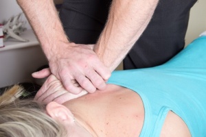
Spinal manipulation (SM) can improve function and reduce spinal disability.  SM also provides pain relief for many disorders such as back pain and neck pain.   Pain induces changes in both the central and peripheral nervous systems.  The mechanisms by which SM alters musculoskeletal pain are still not completely known.  Current evidence however suggests that SM is associated with neurophysiological responses including rapid hypoalgesia with simultaneous sympathetic and motor system excitation.  Animal studies have shown that analgesia provided by joint mobilization involves serotonin and noradrenaline receptors in the spinal cord.

A new investigation sought to determine the response of several other biochemical markers of pain and stress to SM.  Specifically, three neuropeptides (neurotensin, oxytocin, orexin A) and a glucorticoid hormone (cortisol) were studied.  The authors note that the neuropeptides have been associated with hypoalgesia and pain modulation and that cortisol plays an analgesic role in the stress response.  Recent theories have suggested that chronic pain could be partially maintained in a facilitated state due to maladaptive responses in the presence of recurrent stressful situations.  To date there is a lack of studies analyzing these specific biomarkers in relation to SM.

The purpose of this study was to determine the effect of cervical or thoracic manipulation on neurotensin (NT), oxytocin, orexin A, and cortisol levels.  Experimenters examined both spinal regions because they thought there may be a difference in anti-nociceptive effect between the cervical spine and thoracic spine.

Participants included graduate students from Spain.  All subjects were asymptomatic and were excluded if there was a contraindication to manipulation, history of whiplash or surgery, pain in the last month, SM in the last 2 months.  Thirty asymptomatic subjects were randomly divided into 3 groups: cervical manipulation (n = 10), thoracic manipulation (n = 10), and non-manipulation (control) (n = 10).  Although it is not explicitly stated in the article, I presume the manipulations were performed by physical therapists since the lead authors were PT’s.  Manipulations consisted of supine ‘anterior’ thoracic spine manipulations, and rotary type cervical manipulations.  Blood samples were extracted before, immediately after, and 2 hours after each intervention by way of venipuncture of the cephalic vein. Neurotensin, oxytocin, and orexin A were determined in plasma using enzyme-linked immuno assay. Cortisol was measured by microparticulate enzyme immuno assay in serum samples.

Results

Neurotensin (NT)

  • Statistically significant increases in neurotensin occurred in both the thoracic and cervical manipulation groups compared to controls post-intervention with the greatest increase occurring immediately following manipulation
  • Cervical spine manipulation produced a slightly larger increase in neurotensin

Orexin A

  • No statistically significant changes were noted in orexin A levels following treatment

Oxytocin

  • Statistically significant increases in oxytocin occurred in both the thoracic and cervical manipulation groups compared to controls post-intervention with the greatest increase occurring immediately following manipulation
  • Cervical spine manipulation produced a significantly larger increase in oxytocin compared to thoracic manipulation

Cortisol

  • A significant increase in cortisol occurred in the cervical manipulation group compared to controls and the thoracic manipulation group immediately post-intervention
  • However, a significant decrease in cortisol was found at 2 hrs post intervention in the thoracic SM group compared with pre-intervention values
  • A non-significant decrease in cortisol was found also found at 2 hrs post intervention in the cervical SM group compared with pre-intervention values


Discussion

NT is an endogenous peptide with broad spectrum of central and peripheral activities, including modulation of pain signal transmission and perception. NT behaves as a neurotransmitter in the brain and as a hormone in the gut.  Because of its association with a wide variety of neurotransmitters, NT has been implicated in the pathophysiology of several CNS disorders such as schizophrenia, drug abuse, Parkinson’s disease (PD), pain, central control of blood pressure, eating disorders, as well as, cancer and inflammation. Note that the antinociceptive effects of NT are independent from opioid antinociception.

Increased oxytocin following SM could be partly responsible for the analgesic effect linked to manual therapy techniques due to the activation of descending pain-inhibitory pathways.

Cortisol is a potent anti-inflammatory that functions to mobilize glucose reserves for energy and modulate inflammation. Ultimately, a prolonged or exaggerated stress response may perpetuate cortisol dysfunction, widespread inflammation, and pain.  SM in this study led to an immediate increase in cortisol followed by a significant 2 hour decrease in levels with thoracic manipulation and a decrease in 2 hr levels with cervical manipulation.


Key Points

  • SM can modify several biochemical markers of pain and stress
  • These findings suggest that descending inhibitory pathway mechanisms may be involved in the physiological effects that follow SM
  • The effect size for the cervical manipulation group was larger than that for the thoracic manipulation group suggesting an increase in the activation of the possible descending inhibitory pathway mechanisms after cervical manipulation compared to thoracic manipulation

Reference: Plaza-Manzano G, Molina-Ortega F, Lomas-Vega R, Martínez-Amat A, Achalandabaso A, Hita-Contreras F. Changes in biochemical markers of pain perception and stress response after spinal manipulation. J Orthop Sports Phys Ther. 2014 Apr;44(4):231-9.

Learn the basics about Evidence Based Practice for chiropractors and some quick strategies to search the scientific literature regarding chiropractic.  Watch in HD for the best viewing experience.

Dean Smith, DC, PhDChiropractors, how do you take all of the current chiropractic research and develop an effective evidence based practice strategy? I will be joining Dr. Stu Hoffman and ChiroSecure next Tuesday, June 21st for The Importance of an Evidence Based Practice.

At this event you will learn:

  • What is an Evidence Based Practice?
  • The Importance of an Evidence Based Practice
  • Barriers to the Evidence Based Practice

Dr. Dean Smith is a practicing chiropractor and has published over 40 peer reviewed scientific articles and conference publications.  Dr. Smith will be presenting what leading chiropractic researchers are saying about their own research (from the chiropractic science podcast).  We will also discuss strategies to incorporate the latest science into your practice.

So mark your calendars for the following:

WHO SHOULD ATTEND:  All chiropractors, associates and staff

WHEN: Tuesday, June 21st, 2016, 2:00 PM EST

WHERE: http://www.chirosecure.com/live/

LENGTH: 30 to 40 minutes

HOW TO SIGN-UP: JUST SHOW UP NO OPT-IN

COST TO ATTEND: NONE, COURTESY OF CHIROSECURE*

REPLAY: For ChiroSecure Concierge Members

PLEASE NOTE: We will be providing valuable notes to every attendee of the Live Event.

CHIROSECURE CONCIERGE SERVICE: Sign up for our Concierge Service and we will automatically send you the video replay, the notes and the transcripts for all our Live Events without ever having to show up again. http://www.chirosecure.com/concierge-service/

*Supporting the Chiropractic community for over 25 years,  ChiroSecure’s Live Events educate and support you, the practitioner, by making sure you have the information you need to protect you, your practice and your future.


18448850_xxl
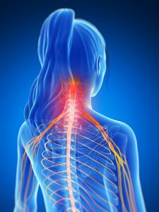
The application of spinal manipulative therapy (SMT) is a cost-effective and widely recognized manual intervention used by a variety of health care professionals in the management of musculoskeletal pain. A growing body of scientific evidence supports the use of SMT for the treatment of a broad range of musculoskeletal disorders citing short-term antinociceptive (pain-relieving) effects and restoration of normal joint mechanics.

Last year, about this time, I wrote about a systematic review that found spinal manipulation therapy (SMT) has a pain reducing effect as measured by pressure pain thresholds (PPT).  Additionally, the effect of SMT on pain reduction was statistically significant at remote locations (for example, adjusting the neck yielded reduction in pain at the elbow).

A new study has emerged in the scientific literature that advances our understanding of the topic (Srbely et al, 2013).  The authors note that although the pathophysiology of myofascial pain syndrome (MPS) is still unclear, research suggests that myofascial trigger points (MTPs) play an fundamental role in the generation and clinical manifestation of MPS.  However, it is currently unknown if the antinociceptive effects of SMT in myofascial tissues are manifest predominantly via regional or general mechanisms, or a combination of both. A study was needed to specifically investigate the hypothesis that SMT evokes robust antinociceptive effects in MTPs preferentially located within neurosegmentally linked myofascial tissues.

Srbely et al conducted the study through the University of Guelph.  The study was a single session, single blinded, randomized controlled intervention.  The primary inclusion criterion was the presence of a clinically identifiable MTP locus (active or latent) within the right infraspinatus and right gluteus medius muscles.  The primary diagnostic criterion used to clinically identify the trigger point locus was ‘a palpable hyperirritable nodule nested within a taut band of skeletal muscle; sustained ischemic pressure over the trigger point locus elicited a dull achy regional pain or discomfort.’  Exclusion criteria encompassed conditions that would affect normal somatosensory processing.

Thirty-six participants qualified for the study and were randomly assigned to test or control groups.  Two chiropractors saw participants at an urban outpatient clinic. One chiropractor performed the history, exam and manipulations while the other chiropractor (blinded to treatment allocation) detected the trigger points and measured all PPTs. The primary outcome was PPT values from infraspinatus and gluteus medius muscles.  The infraspinatus was chosen due to its innervation from the manipulated segment (C5-6). The gluteus medius acted as a regional control point (L4-S1 innervation).

PPT was measured with a force gauge (Newtons) over the trigger point locus (infraspinatus, gluteus medius) and was defined as the force necessary to elicit the onset of a deep dull achy local discomfort and/or referred pain. Measurements were taken at 1,5, 10, and 15 minutes postintervention.  In order to specifically compare regional antinociceptive effects between intervention groups, the authors also calculated the PPT difference (PPTdiff) between infraspinatus and gluteus medius trigger points at each time interval within each participant. Participants received a rotary type manipulation to the C5-6 segment in a supine posture.   Additionally, a drop piece mechanism was used to aid in the high velocity low amplitude thrust.  Control participants received a sham manipulation.  The sham consisted of rotating the neck of the participant, supporting the neck of the participant with the clinician’s forearm under the headpiece and a thrust of the forearm into the headpiece.  It is noted that the contact hand did not thrust and did not create ‘a real manipulation’ of any segment.

Results:

  • there was a significantly increased PPT threshold for infraspinatus trigger points in treated participants compared to controls at all time intervals beyond baseline
  • there was a significantly increased PPT threshold for infraspinatus compared to gluteus medius before and after manipulation at all time intervals beyond baseline
  • no significant differences in PPT scores were observed at any time interval when comparing test gluteus medius, control infraspinatus, and control gluteus medius groups
  • there were significant increases in PPTdiff in the test group vs controls at all time intervals beyond baseline

Key Findings:

  • This study suggests that SMT evokes statistically significant short-term increases in PPT in segmentally related myofascial tissues in young adults
  • Decreased pressure sensitivity (increased PPT score) was observed at all time intervals beyond baseline within neurologically linked infraspinatus muscle after real, but not sham, manipulation
  • The peak antinociceptive effect was measured as a 36% decrease in pressure sensitivity from baseline values and was recorded at 5 minutes postSMT

So, what does this study tell us?  It suggests that SMT evokes robust regional antinociceptive effects in myofascial tissues.  It also provides important evidence to support further research into the potential benefit and role of SMT in the management of chronic widespread pain syndromes  including myofascial pain, and fibromyalgia.

Reference: Srbely JZ, Vernon H, Lee D, Polgar M. Immediate effects of spinal manipulative therapy on regional antinociceptive effects in myofascial tissues in healthy young adults. J Manipulative Physiol Ther. 2013 Jul-Aug;36(6):333-41.


pregnantMusculoskeletal pain in pregnant women is common and is frequently viewed as short-lived and temporary.  Most women report either low back pain (LBP) or pelvic pain (PP) during pregnancy and up to 40% of patients report pain during the 18 months after delivery.  Pelvic pain in pregnant women is a health care challenge in which moderate and severe pain develops rather early and has important implications for society.  These pain syndromes increase sick leave and impair general level of function during pregnancy.

Previous research has evaluated different treatments to reduce morbidity in women with LBP and/or PP during pregnancy including prescription exercise, manual manipulation, education, acupuncture, and pelvic belts amongst others.

The authors of this study conducted a prospective, randomized, masked clinical trial (including 169 women) to test the hypothesis that a multimodal approach (MOM) involving a chiropractor including manual therapy, exercise, and education for LBP/PP in pregnant women is superior to standard obstetric care (STOB) for reducing pain, impairment, and disability in the antepartum period.

Patients from this study were recruited from 3 university affiliated practices (Washington University, St. Louis, MO).  Patients were between 15-45 years old with a single fetus from 24-28 weeks’ gestation.  Patients were evaluated by their obstetric provider for LBP, PP or both.  Exclusion criteria included acute inflammatory disease, acute infectious disease, chronic back pain for greater than 8 weeks before pregnancy, a mental health disorder, back pain from visceral disease, ongoing treatment for previous back pain, peripheral vascular disease, substance abuse, or litigation pending from back pain.

A single, masked chiropractic specialist conducted the baseline evaluation (BE) with eligible volunteers before randomization.  Randomization of subjects across to the STOB group or the STOB plus multimodal musculoskeletal and obstetric treatment (MOM) group was achieved across all 3 sites.

Subjective and objective measures were collected at baseline evaluation (24-28 weeks’ gestation) with follow-up at 33 weeks’ gestation. Three questionnaires including the numerical rating scale, Quebec task force disability questionnaire (QDQ) and personal pain history (PPH) were obtained.  Physical assessments included the straight leg raise (SLR), posterior PP provocation test, active SLR, and long dorsal ligament test.

Patients in the both the STOB and MOM groups selected their own obstetric provider.   In addition to obstetric care, the MOM group had weekly visits with a chiropractor until 33 weeks’ gestation who provided education, manual therapy, and lower back and pelvic stabilization exercises.  Patients were reassured the pain experienced was not likely pathologic and that activation of joints and muscle through exercise would likely improve symptoms and signs without posing risk to the patient or her fetus.  Joint mobilization involved gently moving hypomobile joints in their restricted directions to help restore proper range of motion.  The gluteus maximus, gluteus medius, quadratus lumborum, abdominal wall, and intrinsic spine muscles were targeted in the quadruped, supine, or side-lying positions.

Key findings of this study were:

  • the MOM group (involving chiropractor) had a significant reduction in pain on 7 indices (NRS, QDQ, SLR(left), active SLR, long dorsal ligament test, PPH – leg and shoulders)
  • the STOB group had a significant increase in pain on 5 indices and only 1 improvement (PPH -leg)
  • The MOM group reported significantly less trouble sleeping at 33 weeks’ gestation than the STOB group
  • No adverse events were reported in either group

In summary, including chiropractic interventions with standard obstetric care for low back and pelvic pain in mid pregnancy benefits patients more than standard obstetric care alone. The benefits derived are both subjective and objective. Patients perceived less pain and disability and an overall global improvement in daily activities. Their physical examinations revealed improved range of motion, stability, and less irritation at the lumbar and pelvic joints.

Reference:

George JW, Skaggs CD, Thompson PA, Nelson DM, Gavard JA, Gross GA. A randomized controlled trial comparing a multimodal intervention and standard obstetrics care for low back and pelvic pain in pregnancy. Am J Obstet Gynecol. 2013 Apr;208(4):295.e1-7.


35856944_s
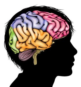
Research on chiropractic spinal manipulation (CSM) has been conducted extensively worldwide, and its efficacy on musculoskeletal symptoms has been well documented.  Previous studies have documented potential relationships between spinal dysfunction and the autonomic nervous system and that chiropractic treatment affects the autonomic nervous system. The authors of this study hypothesized that CSM might induce metabolic changes in brain regions associated with autonomic nervous system functions as assessed with positron emission tomography (PET).  Positron emission tomography is a nuclear medicine imaging technique that allows quantification of cellular and molecular processes in humans such as cerebral glucose metabolism which is thought to reflect regional neuronal activities.

Participants were men between the ages of 20-40 who had neck pain and shoulder stiffness.

A crossover study design was used such that subjects served as their own controls to compare their resting brain activity to their brain activity following chiropractic manipulation.  Half of the participants completed the control condition first while the other half completed the chiropractic condition first.  The participants came back sometime between 1 and 6 weeks later to complete their remainder condition.  Chiropractic consisted of a single Activator Methods assessment and treatment session by a chiropractor lasting 20 minutes.  The control condition consisted of 20 minutes of rest.  Immediately after each condition, 18F-labeled fluorodeoxyglucose (FDG) was injected.  FDG is an excellent imaging marker of brain metabolism (glucose consumption).  PET scanning followed administration of FDG.

Additional outcome measures included pain (VAS), Stress Response Scale (SRS-18) and European Organization for Research and Treatment of Cancer Quality of Life Questionnaire-Core 30 (EORTC QLQ-C30), trapezius muscle tone, and salivary amylase.

Results of the PET image analysis showed statistically significant changes in regional cerebral metabolism between rest and treatment conditions.  With chiropractic treatment, increased glucose metabolism was observed in the inferior prefrontal cortex, anterior cingulate cortex,  middle temporal gyrus; decreased glucose metabolism was observed in the cerebellar vermis and visual association cortex.  Reduced metabolism in the cerebellar vermis may be related to reductions is pain, mental stress, muscle tone and sympathetic tone.  Activation of the anterior cingulated cortex and inferior prefrontal cortex may arise from sympathetic relaxation.

The mean SRS-18 and EORTC QLQ-C30 scores were significantly lower in the treatment condition indicating improved stress response and improved quality of life. Mean VAS pain score comparison was significantly improved with treatment.  Additionally, measurement of trapezius muscle tone and salivary amylase showed significant reduction with chiropractic suggesting improved sympathetic relaxation.


Reference: Tashiro M, Ogura T, Masud M, Watanuki S, Shibuya K, Yamaguchi K, Itoh M, Fukuda H, Yanai K. Cerebral metabolic changes in men after chiropractic spinal manipulation for neck pain. Altern Ther Health Med. 2011 Nov-Dec;17(6):12-7.


1005989_highIn a recent study, manual manipulation by a chiropractor led to greater short term reductions in self-reported pain and disability than manual assisted manipulation (Activator) or usual medical care by a physical medicine and rehabilitation specialist.

Low back pain (LBP) is an extremely common presenting complaint that occurs in greater than 80% of people. Chiropractors care for patients who have no symptoms and those who have symptoms.  Chiropractic has been used as a treatment for those with lower back pain but evidence is mixed with some reviews finding no advantage of chiropractic spinal manipulation therapy (SMT) compared to other treatments while some guidelines find moderate effectiveness of chiropractic care for back pain.  Research has demonstrated that chiropractic care in addition to standard medical care improves pain and disability scores, and in another study a subgroup of patients with acute nonspecific LBP – spinal manipulation was significantly better than nonsteroidal anti-inflammatory drug diclofenac and clinically superior to placebo (Spine 2013; 38:540-548).  The study reviewed here sought to compare the effectiveness of manual thrust manipulation (MTM) and manual assisted manipulation (MAM), to usual medical care (UMC) for the treatment of acute and subacute LBP.

Methods:

This study was a prospective, randomized controlled trial evaluating the comparative effectiveness of manual and mechanical spinal manipulation to usual medical care for the treatment of acute and subacute LBP.  Participants were at least 18 years old and had a new LBP episode within the previous 3 months.  They also were required to have a minimum level of self-rated pain of 3 out of 10 and minimum disability rating of 20 out of 100. Exclusions included: chronic LBP (greater than 3 months duration), previous treatment for the current episode, radicular signs/symptoms, contraindications to SMT, current use of prescription pain medicine.

Participants and treating clinicians were not blinded to treatment allocation but the principal investigator was blinded to treatment assignment and had no interaction with participants.

The study interventions consisted of:

  1. Manual thrust manipulation (MTM) – high velocity low amplitude thrust delivered by a chiropractor to the lower thoracic, lumbar and SI joints in the side posture position as deemed necessary
  1. Mechanical-assisted manipulation (MAM) – certified Activator Methods chiropractor delivered MAM in the prone position to the lower thoracic, lumbar and SI joints as deemed necessary
  1. Usual medical care (UMC) – participants were seen by a board certified physical medicine and rehabilitation medical doctor and prescribed over the counter analgesic and NSAID medications, given advice to stay active and avoid bed rest

All groups had a 4 week course of care.  All groups received an educational booklet describing proper posture and movements during activities of daily living. Both manipulation groups had 8 visits (2 per week x 4 wks).  The UMC group had 3 visits (initial, at 2 weeks and at 4 weeks).  Following the 4 week assessment, participants were free to pursue rehabilitation or manipulation.

The primary outcome was the Oswestry LBP Disability Index (OSW) and this index provides a valid and reliable way to assess functional impairment.  Pain intensity ratings were also collected using the mean of current pain, worst pain in 24 hours and average pain during last week.  Outcomes were assessed at baseline, 4 weeks, 3 months and 6 months. Other outcomes were physical examination, fear avoidance questionnaire, and treatment credibility-expectation questionnaire.

Participants with at least 30% or 50% reductions in an outcome measure were considered to be ‘responders’ and had moderate or substantial improvement respectively.

Results:

  • No adverse events were reported

Longitudinal Analysis:

  • For disability, no statistically significant differences were found between groups
  • For pain, MTM yielded lower pain scores compared to MAM and UMC
  • For pain, there were no significant differences between MAM and UMC

Responder Analysis:

  • 76% of MTM group achieved at least 30% reduction in disability compared with about 50% of MAM and 50% of the UMC groups (MAM not significantly different from UMC)
  • 50% of MTM group achieved at least a 50% reduction compared with 16% of the MAM and 39% of the UMC groups (MAM was significantly worse than UMC in this outcome)
  • 94% of MTM achieved greater than 30% reduction in pain compared with 69% of MAM and 56% of UMC
  • 76% of MTM achieved greater than 50% reduction in pain compared to 47% of MAM and 41% of UMC (MAM not significantly different from UMC)

Key Points:

  • Manual thrust manipulation by a chiropractor led to greater short term reductions in self-reported pain and disability than manual assisted manipulation (Activator) or usual medical care by a physical medicine and rehabilitation specialist
  • The benefits seen at the end of 4 weeks of care was no longer statistically significant at 3 or 6 months
  • MTM should be considered as an effective short term treatment option for patients with acute and subacute LBP
  • Significantly more patients in the MTM group achieved moderate or substantial reductions in disability and pain scores
  • These results contradict assumptions of therapeutic similarity between manual thrust and mechanical-assisted manipulation

Reference: Schneider M, Haas M, Glick R, Stevans J, Landsittel D. Comparison of spinal manipulation methods and usual medical care for acute and subacute low back pain: a randomized clinical trial. Spine (Phila Pa 1976). 2015 Feb 15;40(4):209-17.

Hip Arthritis
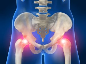
Chiropractic care holds potential value for the treatment of a variety of limb conditions.  For patients with osteoarthritis (OA) of the hip, a combined intervention of manual therapy provided by a chiropractor and patient education was more effective than a minimal control intervention.

A recent article by Poulsen et al (2013) contributes to our understanding of the literature regarding chiropractic and lower extremity conditions – particularly hip osteoarthritis. Hip osteoarthritis (OA) is a common joint disease and when symptomatic can have significant impact on regular daily activities.  Recently, hip OA has been linked to higher mortality rates.  In end stage hip OA, joint replacement surgery is an appropriate and cost-effective treatment but a long-term cohort study has documented that only 20% of patients with radiographic hip OA have had surgery 11-28 years after the initial diagnosis.  Therefore, non-surgical interventions with documented effectiveness become essential for patients who do not need, or choose not to have surgery.

Although guidelines recommend patient education (PE) programs as a core intervention, systematic reviews are contradictory in conclusions regarding their effectiveness on pain and function in hip OA. Manual therapy (MT) has been proposed as an adjunct intervention to exercise for patients with hip OA but evidence is based on a single randomized clinical trial (RCT).  The authors of the current study realized this gap in the literature and decided to investigate the effectiveness of a patient education (PE) program with or without the added effect of manual therapy (MT) compared to a minimal control intervention (MCI).

The design of this study was a single-center proof-of-principle three-arm parallel group RCT.  Inclusion criteria were: Unilateral hip pain >3 months’ duration, age 40-80 years, radiographic hip OA defined as minimal joint space width (JSW) measurement <2.00 mm or a side difference in minimal JSW >10%, and, ability to speak and read Danish.  The study took place at the Department of Orthopaedic Surgery and Traumatology, Odense University Hospital, Denmark.

During the first 2 months of recruitment, 3 exclusion criteria were added to the original criteria: patients who had had MT within the previous twelve months; patients who rated their pain severity as 1 or 2 on the numerical rating scale (NRS); patients with polyarthritis.

The 3 groups in the study were: 1) MCI; 2) PE; 3) PE + MT.

For the MCI group, a nurse provided written advice on a home stretching program derived from the PE program together with 5-10-min instruction.

The PE group, originally termed ‘Hip School’ was taught by a physiotherapist with 11 years experience. The PE program included two individual sessions and three group sessions.

In the combined PE and MT group, manual therapy was administered by a chiropractor with 20 years of clinical experience. MT was scheduled twice a week for the 6-week intervention period and treatment was individualized to each patient depending on examination findings. MT consisted of: trigger point release therapy (TPPR), muscular stretching by muscle energy technique (MET) and joint manipulation.

The primary outcome was pain severity rated on an 11-box NRS, measured after 6 weeks of intervention. Patients were asked to rate the worst pain experienced during the previous week. Secondary outcome measures were the Hip Disability and Osteoarthritis Outcome Score (HOOS) ranging from 0-100, worst to best; patients’ perceived global effect of interventions, percentage in each group having classified themselves as improved; passive hip range of motion (ROM); use of pain medication at 12 months and hip replacement surgery within the 12 month follow-up period.

Results:

  • A total of 111 patients were included in the analyses at the primary end point at 6 weeks
  • In the combined group (PE + MT), a clinically relevant reduction in pain severity compared to the MCI of 1.90 points was achieved
  • Effect size (Cohen’s d) for the PE + MT minus the MCI was 0.92 (large effect)
  • The number of patients in each group experiencing pain reduction of at least 25% from baseline to 6 weeks was PE = 8, PE +MT = 21 and MCI = 7
  • Number needed to treat for PE + MT was 3
  • No difference was found between the PE and MCI groups
  • At 12 months, not including patients receiving hip surgery the statistically significant difference favoring PE + MT was maintained
  • All HOOS (pain, symptoms, ADLs, Sport/Rec, QOL) subscales demonstrated clinically relevant and statistically significant superiority, p < 0.05 for the PE + MT group when compared to the MCI group
  • Mean differences between PE and MCI were small (range 4 to 1) and not statistically significant, p > 0.05
  • Effect sizes for HOOS subscales for PE + MT minus MCI ranged between 0.75 and 1.08
  • No changes in hip ROM noted between groups

Key Findings:

  • For primary care patients with OA of the hip, a combined intervention of manual therapy provided by a chiropractor and patient education was more effective than a minimal control intervention
  • Note that patient education alone was not superior to the minimal control intervention

So, what does this study tell us?  This trial demonstrated clinical and statistically significant improvements in pain, symptoms and disability for a combined intervention consisting of manual therapy provided by a chiropractor and patient education when compared to a minimal control intervention including home stretching.


Reference: Poulsen E, Hartvigsen J, Christensen HW, Roos EM, Vach W, Overgaard S. Patient education with or without manual therapy compared to a control group in patients  with osteoarthritis of the hip. A proof-of-principle three-arm parallel group randomized clinical trial. Osteoarthritis Cartilage. 2013 Oct;21(10):1494-503.

ChiropracticOver the last decade, research has demonstrated that spinal manipulation can change various aspects of nervous system function, including muscle reflexes, cognitive processing, reaction time, and the speed at which the brain processes information. One research group from New Zealand (Haavik et al) has hypothesized that the joint dysfunction part of the chiropractic clinical construct, the vertebral subluxation, results in altered afferent input to the central nervous system (CNS) that modifies the way in which the CNS processes and integrates all subsequent sensory input. This processing (i.e., sensorimotor integration) is a central nervous system (CNS) function that appears most vulnerable to altered inputs.

Many studies show that chiropractic adjustments result in changes to sensorimotor integration within the central nervous system. Do these changes correlate with beneficial clinical outcomes?  That is not completely determined yet. It is also not clear whether the changes seen after adjustments are due to the correction of vertebral subluxation, therefore normalizing aberrant afferent input to the CNS, or are they merely due to afferent influx associated with the thrusting into the spine? These questions remain to be answered. The level of CNS involvement and the exact mechanisms underlying these neural adaptations following chiropractic adjustments remain unclear.

This new study reported here sought to investigate possible neural plastic changes with spinal manipulation by measuring H-reflexes and V-waves.  The H-reflex is an electrically evoked response that operates via the same neuronal circuitry as stretch reflexes.  The H (Hoffmann) reflex may be useful to assess motoneuron excitability in vivo while also reflecting presynaptic inhibition of Ia afferent synapses.  The so-called V-wave, which is an electrophysiological variant of the H-reflex, can be recorded during maximal voluntary motor contractions. The elicited V-wave response may be used to reflect the level of efferent neural drive from spinal α-motoneurons during maximal voluntary contraction (MVC).

While several previous studies have shown a decrease in the H-reflex indicating a transient attenuation of motoneuronal activity of the lumbosacral spine in both asymptomatic subjects and low back pain patients, new advances in data collection and processing have occurred since then. The purpose of this study was to take advantage of these recent technical and methodological discoveries related to the H-reflex and V-waves and explore what effect, if any, spinal manipulation of vertebral subluxations will have on them.

Methods:

Two studies were included in the paper.  All participants were men, between the ages of 18 and 40 and were required to have evidence of spinal dysfunction and a previous history of subclinical pain, but absence of degenerative conditions of the spine or known contraindications to spinal manipulation.  Instrumentation included: 1) surface EMG to record the (SEMG) activity of the soleus muscle (SOL) of the right leg; 2) electrical stimulation producing the H-, M-, and V-waves of the SOL muscle by stimulation of the tibial nerve and; 3) force recordings performed using a strain gauge attached to a custom-made ankle brace, while the subject performed maximum voluntary ankle dorsiflexion contractions.

During study one, ten subjects attended two sessions each, the control and the experimental (spinal manipulation) session. A second study was added wherein a group of eight participants attended two more sessions each, where only force was measured. The order of these sessions were randomized and at least 1 week separated the sessions.  All experiments were performed on the right leg, while the volunteers comfortably lay prone on a massage table with their right leg firmly strapped to the table with Velcro.  The following measures were collected pre and post interventions: SEMG signals during MVC; H-and M-recruitment curves; H-reflex area under curve normalized to Mmax (Harea/Mmax), H-reflex threshold, V-wave normalized to Mmax (V/Mmax), M-wave slope, H-reflex slope and the mean power frequency (MPF) of a fast Fourier transform (FFT) of the SEMG during MVC.

The entire spine and sacroiliac joints were assessed for segmental dysfunction (vertebral subluxation) and adjusted where deemed necessary by a registered chiropractor with at least 10-years clinical experience using high-velocity, low-amplitude techniques.  The control condition involved passive and active movements of the subject’s head, spine, and body into the manipulation setup positions but without performing the adjustment.

Results:

  • the threshold to elicit the H-reflex significantly decreased by 8.5% in the spinal manipulation group
  • the SEMGs showed a significant drop in the power spectrum after controls but there was no fatigue demonstrated in the power spectrum after spinal manipulation
  • for study 1: maximal voluntary contraction as determined by SEMG increased significantly by 59.5% after spinal manipulation and decreased significantly by 13.3% after control
  • for study 2: maximal voluntary contraction increased significantly by 16.1% after spinal manipulation and decreased significantly by 11.4% after control
  • the V-wave amplitude (V/Mmax ratio) increased significantly by 45% after spinal manipulation and reduced significantly by 23% after control

Key Points

  • this study is the first to indicate that chiropractic adjustments can induce significant changes in the net excitability for the low-threshold motor units/and or alters the synaptic efficacy of the Ia synapse
  • the improvements in maximal voluntary contraction following spinal manipulation are likely attributed to the increased descending drive and/or modulation in afferents
  • spinal manipulation prevents fatigue
  • these results suggest that spinal manipulation may be indicated as part of the treatment for the patients who have lost tonus of their muscle and/or are recovering from muscle dysfunction such as stroke or orthopedic operations
  • these findings will also be of interest to athletes and perhaps the general public


Reference: Niazi IK, Türker KS, Flavel S, Kinget M, Duehr J, Haavik H. Changes in H-reflex and V-waves following spinal manipulation. Exp Brain Res. 2015 Apr;233(4):1165-73. doi: 10.1007/s00221-014-4193-5. Epub 2015 Jan 13. PubMed